Barium »
PDB 7awn-9b3g »
7cmj »
Barium in PDB 7cmj: Crystal Structure of L.Donovani Hypoxanthine-Guanine Phosphoribosyl Transferase (Hgprt)
Enzymatic activity of Crystal Structure of L.Donovani Hypoxanthine-Guanine Phosphoribosyl Transferase (Hgprt)
All present enzymatic activity of Crystal Structure of L.Donovani Hypoxanthine-Guanine Phosphoribosyl Transferase (Hgprt):
2.4.2.8;
2.4.2.8;
Protein crystallography data
The structure of Crystal Structure of L.Donovani Hypoxanthine-Guanine Phosphoribosyl Transferase (Hgprt), PDB code: 7cmj
was solved by
P.S.Parihar,
J.V.Pratap,
with X-Ray Crystallography technique. A brief refinement statistics is given in the table below:
| Resolution Low / High (Å) | 44.37 / 2.76 |
| Space group | P 65 2 2 |
| Cell size a, b, c (Å), α, β, γ (°) | 80.8, 80.8, 344.321, 90, 90, 120 |
| R / Rfree (%) | 18.1 / 22.5 |
Other elements in 7cmj:
The structure of Crystal Structure of L.Donovani Hypoxanthine-Guanine Phosphoribosyl Transferase (Hgprt) also contains other interesting chemical elements:
| Magnesium | (Mg) | 2 atoms |
| Chlorine | (Cl) | 2 atoms |
Barium Binding Sites:
The binding sites of Barium atom in the Crystal Structure of L.Donovani Hypoxanthine-Guanine Phosphoribosyl Transferase (Hgprt)
(pdb code 7cmj). This binding sites where shown within
5.0 Angstroms radius around Barium atom.
In total 2 binding sites of Barium where determined in the Crystal Structure of L.Donovani Hypoxanthine-Guanine Phosphoribosyl Transferase (Hgprt), PDB code: 7cmj:
Jump to Barium binding site number: 1; 2;
In total 2 binding sites of Barium where determined in the Crystal Structure of L.Donovani Hypoxanthine-Guanine Phosphoribosyl Transferase (Hgprt), PDB code: 7cmj:
Jump to Barium binding site number: 1; 2;
Barium binding site 1 out of 2 in 7cmj
Go back to
Barium binding site 1 out
of 2 in the Crystal Structure of L.Donovani Hypoxanthine-Guanine Phosphoribosyl Transferase (Hgprt)
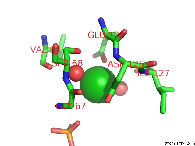
Mono view
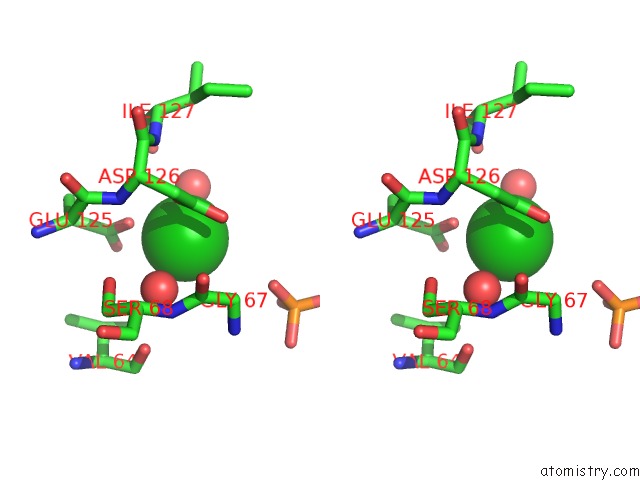
Stereo pair view
Mono view
Stereo pair view
A full contact list of Barium with other atoms in the Ba binding
site number 1 of Crystal Structure of L.Donovani Hypoxanthine-Guanine Phosphoribosyl Transferase (Hgprt) within 5.0Å range:
|
Barium binding site 2 out of 2 in 7cmj
Go back to
Barium binding site 2 out
of 2 in the Crystal Structure of L.Donovani Hypoxanthine-Guanine Phosphoribosyl Transferase (Hgprt)
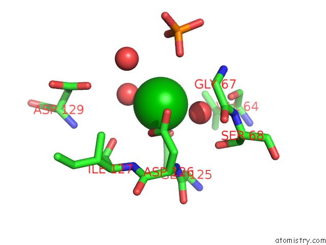
Mono view
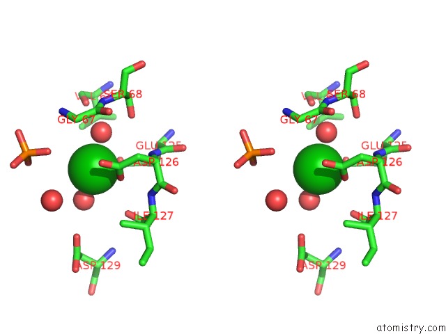
Stereo pair view
Mono view
Stereo pair view
A full contact list of Barium with other atoms in the Ba binding
site number 2 of Crystal Structure of L.Donovani Hypoxanthine-Guanine Phosphoribosyl Transferase (Hgprt) within 5.0Å range:
|
Reference:
P.S.Parihar,
J.V.Pratap.
Crystal Structure of L.Donovani Hypoxanthine-Guanine Phosphoribosyl Transferase (Hgprt) To Be Published.
Page generated: Mon Jul 7 02:49:56 2025
Last articles
Cl in 6DOTCl in 6DNE
Cl in 6DN7
Cl in 6DN3
Cl in 6DMG
Cl in 6DMI
Cl in 6DM1
Cl in 6DM0
Cl in 6DLZ
Cl in 6DLI